Conference Presenters
"DIGITAL WORKFLOW FOR FUNCTIONAL AND AESTHETIC FULL MOUTH REHABILITATION: A CASE REPORT"
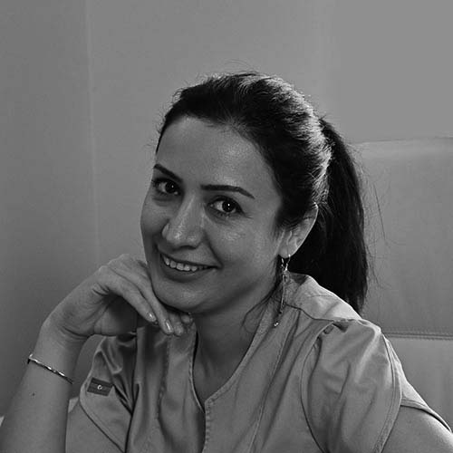
- Dr. Sitara Mammadov (&Dr. Elnur Mammadov)
- MD, ICCMO member
- France

Biography
- Doctor of Medecine option Stomatology (Azerbaïdjan)
- Diploma of Académie d’Occlusion neuromusculaire (ACAEF) (Aix en Provence)
- Membre de l'ICCMO (France)
Abstract
The purpose of this publication is to describe the various stages of managing a patient requiring full rehabilitation of her dental arches, which have undergone significant changes over time, including the loss of a considerable number of teeth. We will present the complete digital workflow that enabled us to carry out this rehabilitation.
The first step involves establishing a diagnosis based on a questionnaire, a clinical examination, and a digital examination protocol: the MAC protocol (following the recommendations of Dr. Combadazou and Professor Destruhaut from the University of Dentistry in Toulouse). This diagnosis should allow us to determine whether the patient is experiencing pain and/or dysfunctions that may be related to the patient’s oral health, or if other postural sensors are involved. Once this diagnosis is established, the search for a therapeutic inter-arch relationship is considered to develop a treatment plan that includes several stages:
**Step 1: Diagnosis and treatment plan**
Impression and digital recording of the therapeutic inter-arch relationship (obtained with K7 and Myomonitor). Recording the range of mandibular movements using the PROSYSTOM for programming the virtual articulator in EXOCAD software. Face scanning using RAYFACE to ensure our prosthetic project aesthetically matches the patient's face. The prosthetic project is created using EXOCAD software. In the case of implant placement, as is the case for our patient, a CBCT is performed to ensure the feasibility of our implant treatment.
**Step 2: Surgery and immediate loading**
Once the aesthetic project is accepted by the patient, guided surgery using XGUIDE allows precise implant placement with minimal tissue damage. After the implants are placed, new digital impressions are taken. To enhance accuracy, given the distortion of digital impressions concerning the full arches, photogrammetry using ICAM is necessary. Immediate loading is carried out using temporary prosthetics.
**Step 3: Placement of the final prosthesis**
After the necessary healing time, the final prosthesis is fabricated.
An occlusion check is performed, and in some cases, the therapeutic CRI requires verification, especially as other postural sensors may influence the mandibular posture after correction, and thus impact occlusion. Final aesthetic adjustments are considered, and the digital fabrication of the final prosthesis is then created and placed in the mouth. A follow-up is conducted to refine dental contacts, recording them digitally using OCCLUSENSE.


